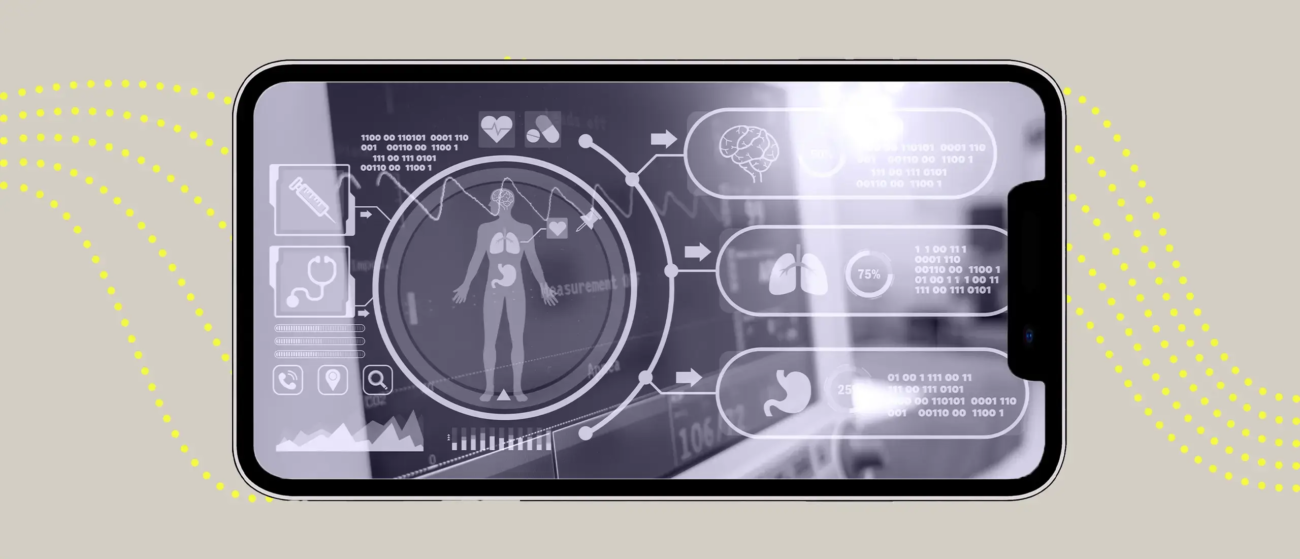
While medical imaging is very well suited for the use of machine learning-based pattern recognition, adoption within healthcare providers is a notoriously slow process due to a lack of trust in AI amongst clinical staff, an unclear economic value proposition which has not been fully proven yet, complex data integration due to siloed and proprietary imaging platforms, not to mention the challenging and uncertain regulatory approval process. In the near future, the algorithm will most likely do initial reading, segmentation and annotation, and any quantification before a radiologist reads the study
The reported accuracy of these algorithms in radiology have varied from 0.56 to 0.99. (3) However, there is no question that this has been improving steadily and the current questions around the use of algorithms in radiology are not so much related to their accuracy and rather their clinical value. Validation of the performance of an algorithm in terms of its accuracy is not equivalent to demonstrating clinical efficacy. Only prospective studies in real world settings can establish that. There have not been many such studies performed to date but the only ones so far are in diabetic neuropathy, wrist fracture, breast cancer histology, colonic polyps, and congenital cataracts.
One area that has been well-established so far is that algorithms can read a scan immediately upon completion and therefore can provide an initial interpretation much faster than a radiologist. In studies to date, this is in order of 150 times faster (1.2 versus 177 seconds.) This can be extremely important in the context of time-sensitive clinical issues, such as stroke, fractures, collapsed lungs, and more conditions.
The benefits of AI in rapidly and accurately reading radiological studies to improve patient care have recently been shown in the context of COVID. Early detection of COVID-19 based on chest CT enables timely treatment of patients and helps control the spread of the disease. In a recent study, an artificial intelligence (AI) system was used for rapid COVID-19 detection based on analysis of chest CTs. The system was developed and evaluated on a large dataset with more than ten thousand CT scans from COVID-19, influenza-A/B, non-viral community acquired pneumonia (CAP) and non-pneumonia subjects. In such a difficult multi-class diagnosis task, the deep convolutional neural network-based system is able to achieve an area under the receiver operating characteristic curve (AUC) of 97.81% for multi-way classification on test cohort of 3,199 scans, AUC of 92.99% and 93.25% on two publicly available datasets, CC-CCII and MosMedData respectively.
In a reader study involving five radiologists, the AI system outperforms all of radiologists in more challenging tasks at a speed of two orders of magnitude above them. In the reader study, the average reading time of radiologists was 6.5 min, while that of AI system was 2.73 s, which can significantly improve the productivity of radiologists.